Free ebooks Library zlib project beylikdüzü escort

Dr. Ivet Bahar receives Kadir Has Outstanding Achievement Award in Turkey

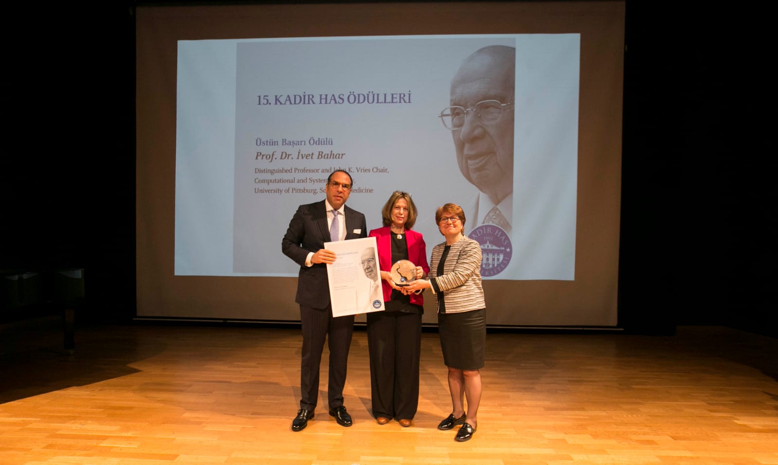
Mr. Nuri Has (left), Dr. Ivet Bahar (middle), and Dr. Sondan Durukanoglu Feyiz (right)

The 15th Annual Kadir Has Awards were held on Friday, March 22nd in Istanbul, Turkey. Member of the Science Academy, Dr. Ivet Bahar, received the Kadir Has “Outstanding Achievement Award” for her contributions to the development of theoretical and computational models for explaining the functional dynamics of biomolecular systems as well as mentoring and teaching a new wave of scientists. She was presented with the award by the Chairman of the Board of Trustees of Kadir Has University, Mr. Nuri Has, the Kadir Has Foundation President, Mr. Can Has, and the President of Kadir Has University, Dr. Sondan Durukanoglu Feyiz.

The Kadir Has Awards seek to recognize the outstanding accomplishments that Turkish scientists have made at the national and international level and to promote people and institutions that have contributed to the development of society.

For more information, please visit http://kadirhasvakfi.org/en/.

Machine learning model can help reduce follow up screenings to 28% of people with benign nodules

A new machine learning model, developed in the Benos lab, can help reduce the unnecessary follow up screenings to people with benign nodules detected in low-dose CT scans by 28%.

Introduction Low-dose CT (LDCT) is currently used in lung cancer screening of high-risk populations for early lung cancer diagnosis. However, 96% of individuals with detected nodules are false positives.

Methods In order to develop an efficient early lung cancer predictor from clinical, demographic and LDCT features, we studied a total of 218 subjects with lung cancer or benign nodules. Probabilistic graphical models (PGMs) were used to integrate demographics, clinical data and LDCT features from 92 subjects (training cohort) from the Pittsburgh Lung Screening Study cohort.

Results Learnt PGMs identified three variables directly (causally) linked to malignant nodules and the largest benign nodule and used them to build the Lung Cancer Causal Model (LCCM), which was validated in a separate cohort of 126 subjects. Nodule and vessel numbers and years since the subject quit smoking were sufficient to discriminate malignant from benign nodules. Comparison with existing predictors in the training and validation cohorts showed that (1) incorporating LDCT scan features greatly enhances predictive accuracy; and (2) LCCM improves cancer detection over existing methods, including the Brock parsimonious model (p<0.001). Notably, the number of surrounding vessels, a feature not previously used in predictive models, significantly improves predictive efficiency. Based on the validation cohort results, LCCM is able to identify 30% of the benign nodules without risk of misclassifying cancer nodules.

Discussion LCCM shows promise as a lung cancer predictor as it is significantly improved over existing models. Validated in a larger, prospective study, it may help reduce unnecessary follow-up visits and procedures.

This is an open access article distributed in accordance with the Creative Commons Attribution Non Commercial (CC BY-NC 4.0) license, which permits others to distribute, remix, adapt, build upon this work non-commercially, and license their derivative works on different terms, provided the original work is properly cited, appropriate credit is given, any changes made indicated, and the use is non-commercial.

Press:

UPMC: https://www.upmc.com/media/news/031219-lung-ca-machine-learning

NPR: https://www.wesa.fm/post/artificial-intelligence-could-reduce-false-positives-lung-cancer-screenings

Vineet K. Raghu


Takis Benos   


      David Wilson


Raghu, …, Benos*, Wilson. Feasibility of lung cancer prediction from low-dose CT scan and smoking factors using causal modelsThorax, 2019

Dr. Jingyu Zhang from Xing’s lab published a first-author paper in PLoS Computational Biology

Spatial clustering and common regulatory elements correlate with coordinated gene expression

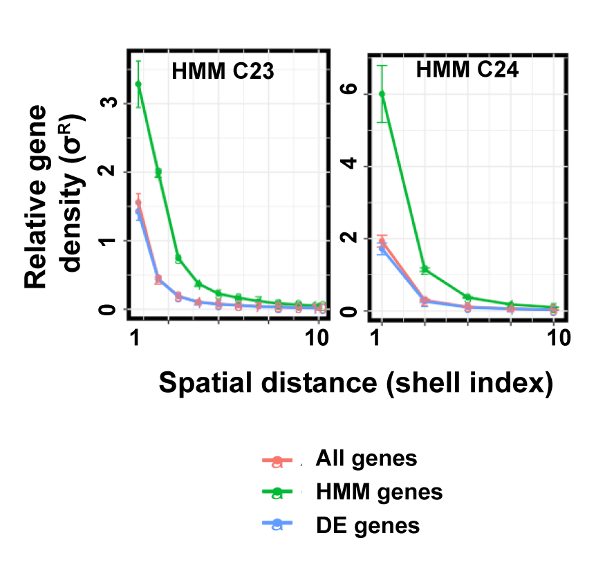
Cellular responses to environmental stimulation are often accompanied by changes in gene expression patterns. Genes are linearly arranged along chromosomal DNA, which folds into a three-dimensional structure. The chromosome structure affects gene expression activities and is regulated by multiple events such as histone modifications and DNA binding of transcription factors. A basic question is how these mechanisms work together to regulate gene expression. In this study, we analyzed temporal gene expression patterns in the context of chromosome structure both in a human cell line under TGF-β treatment and during mouse nervous system development. In both cases, we observed that genes regulated by common transcription factors have an enhanced tendency to be spatially close. Our analysis suggests that spatial co-localization of genes may facilitate the concerted gene expression.

Zhang J, Chen H, Li R, Taft DA, Yao G, Bai F, Xing J. (2019) Spatial clustering and common regulatory elements correlate with coordinated gene expression. PLoS Computational Biology 15(3):e1006786

Dr. Robin Lee to receive collaborative R01 grant with PI Yuan Chang

Title: Role of a Novel Mitotic 4E-BP1 Protein Isoform in Cellular Transformation
PI: Yuan Chang
Co-I: Robin Lee and Patrick Moore

4E-BP1 is the primary gatekeeper for cancer cell cap dependent protein translation. It is directly targeted by mTOR kinase during interphase. We have found that CDK1/CYCB1 substitutes for mTOR during mitosis to phosphorylate 4E-BP1 generating a novel phosphorylation mark at serine (S) 83 that is not present when mTOR phosphorylates 4E-BP1. Unlike other 4E-BP1 phospho-isoforms, phospho-4E-BP1-S83 preferentially localizes to mitotic centrosomes as well as being diffusely distributed in a speckled pattern in the nucleo/cyto-plasm. A mutant form of 4E-BP1 that is unable to be phosphorylated at S83 partially reverses cell transformation caused by the Merkel cell polyomavirus (MCV) small T oncoprotein. This is particularly interesting since discovery of a novel pathway targeted by a tumor virus has always led to discovery of the same pathway being altered in non-infectious cancers. We developed a new phosphospecific antibody to p4E-BP1S83 that allows us to uniquely identify mitosis-related 4E-BP1 phosphorylation and determine its function in cancer cells. With this antibody, we will first survey phosphor-4E-BP-S83 expression in TCGA cancer tissues and anticipate that this approach will be a sensitive measure for activated CDK1 circuits and will provide unique data on cancer-type specific severity. We will next examine the biology of p4E-BP1S83 by identifying S83 phosphospecific effects on mitogenesis and by examining 4E-BP1’s potential role in regulating translation of specific transcripts during mitosis using ribosomal profiling and novel quantitative single cell imaging techniques. Finally, we will generate knock-in mutant mouse models of inactivated and phosphomimetic mitotic 4E-BP1 to determine its role in tumor susceptibility. Our specific aims will advance our fundamental understanding of how a mitosis-specific, hyperphosphorylated form of 4E-BP1 functions in normally cycling cells and how its dysregulation in cancer cells may contribute to human malignancies.

PI: Yuan Chang, MD Co-I: Robin Lee, PhD Co-I: Patrick Moore, MD, MPH